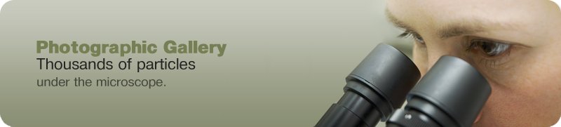
sharing our knowledge.
Photographic gallery.  Thousands of particles under the microscope.

Dispersion Staining, Phase Contrast

G. C Crossmon wrote a paper on the dispersion effects visible with phase contrast illumination in 1949 followed by K. G. Schmidt in 1958. Phase contrast dispersion staining works best with thin particles, such as asbestos fibers. It is extremely sensitive and can easily distiguish asbestos fibers in complex matrices. It has the added advantate of high image resolution and it produces complimentary colors in the same image so that the wavelength of the refractive index match can be more closely ascertained.

Match at 589 nanometers wavelength

Phase Contrast Dispersion Staining

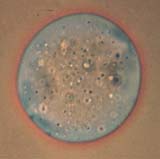
Match at 630 nanometers wavelength, Chrysotile Asbestos Perpendicular to length, Amosite Asbestos Perpedicular to length (650)

Phase Contrast Dispersion Staining Amosite, Phase Contrast Dispersion Staining

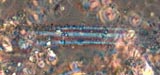
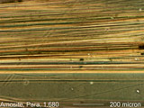
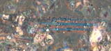
Match at 440 nanometers wavelength, Amosite Asbestos Parallel to length

Phase Contrast Dispersion Staining Amosite, Phase Contrast Dispersion Staining

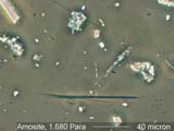
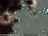
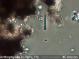
Match at 600 nanometers wavelength, Amosite Asbestos Perpendicular to length, Anthophyllite Asbestos Perpendicular to length (590)

Phase Contrast Dispersion Staining Anthophyllite

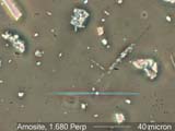
Match at 670 nanometers wavelength,

Phase Contrast Dispersion Staining

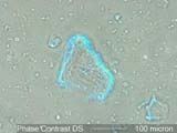
Match at 530 nanometers wavelength, Optical Glass, Chrysotile Asbestos Parallel to length (540)

Phase Contrast Dispersion Staining Phase Contrast Dispersion Staining

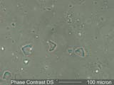
Match at 490 nanometers wavelength, Optical Glass Particles, Anthophyllite Asbestos Parallel to Length (480),

Phase Contrast Dispersion Staining Anthophyllite

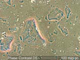
Match at 460 nanometers wavelength

Phase Contrast Dispersion Staining