
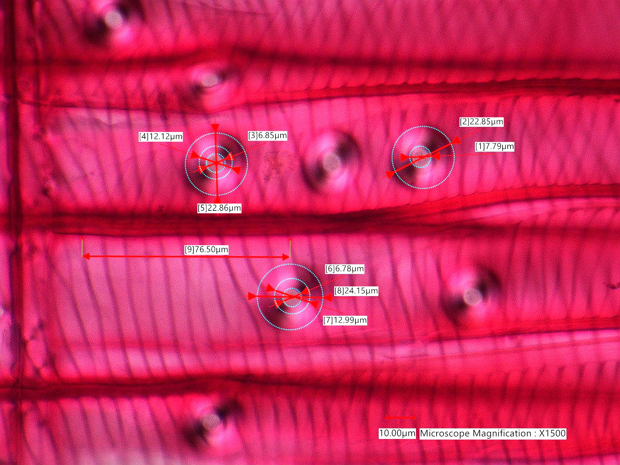
Douglas Fir, Radial-Section
This is an image from a stained standard section of Pseudotsuga menziesii made by John P. Limbach of Ripon, Wisconsin. This shows the helical thickening seen in the vertial tracheids and the inter tracheid pits (pores). The borders of the pits are clearly evident.
Transmitted Off Crossed Linear Polarizing Filters
http://en.wikipedia.org/wiki/Douglas-fir
Definition/Function:
KINGDOM: Plantae DIVISION: Pinophyta CLASS: Pinopsida ORDER: Pinales FAMILY: Pinaceae GENUS: Pseudotsuga SPECIES: menziesiiSignificance in the Environment:
Characteristic Features:
Associated Particles:
References:
Core, H.R., W.A. Cote, and A.C. Day, WOOD STRUCTURE AND IDENTIFICATION, Syracuse Wood Science Series, Vol. 6, 1979.http://en.wikipedia.org/wiki/Douglas-fir
