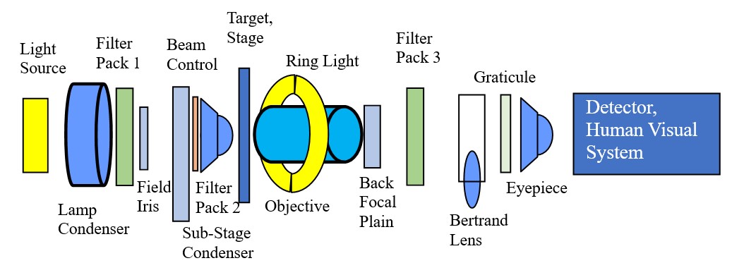
Microscope as a Light Bench
The Light Microscope is basically a optical bench designed for the investigation of the
characteristics of particles. The
first filter pack can control the wavelengths used, type of polarization (linear to
circular), control of tube scatter, and
framing. The second filter pack is at the plane of the substage iris. This is a location
where the "diffracted" beam is in
focus. It is the location for the phase anulus when using phase contrast. It is the
location for Rhineberg filters,
interference system filters, and other filters that change the image by manipulating
parts of the diffraction pattern generated
by the particle and collected by the objective. This location can also be used to
generate different types of dispersion
staining. The angle at which the beam of light strikes the particle can be changed from
parallel to the optical axis of the
microscope to angles greater than the numerical aperture of the objective (about 65
degrees for a dry mount objective).
Transmitted brightfield, transmitted oblique, darkfield oblique, to full darkfield, and
other combinations of illumination
can be controlled at this location. The particle on the stage can be illuminated from
the side by an accessory illuminator
from grazing incidence to oblique reflected darkfield. A ringlight can be attached to
the objective to generate full reflected
darkfield illumination. The angle of the reflected beam can be controlled to some extent
by moving the ringlight up or down
the collar of the objective. The back focal plain of the objective is another
diffraction focal plane. A number of filters
can be place at this location, including the phase plate for phase contrast microscopy
and filters for various types of
interference systems. This is another location where optical stops can be used to
generate dispersion staining. Additional
filters can be placed in the light path above the objective, like compensator plates and
polarizing filters for optical
crystallography. The Bertrand Lens can be inserted to bring the diffraction focal planes
into focus for the observer.
Graticules are placed at the focal plane of the eyepiece to facilitate various
measurement methods applied to the field of
view. The image is then transferred to the human visual system to be refined by a
complex system of built-in algorithms, both
bottom-up and top-down, to create the final perceived image. What is finally seen is
dependent on the skill and knowledge of
the microscopist, just as an infrared spectrum of an organic compound must be
interpreted by one knowledgeable in reading such
data. The human visual system is not a camera.
