
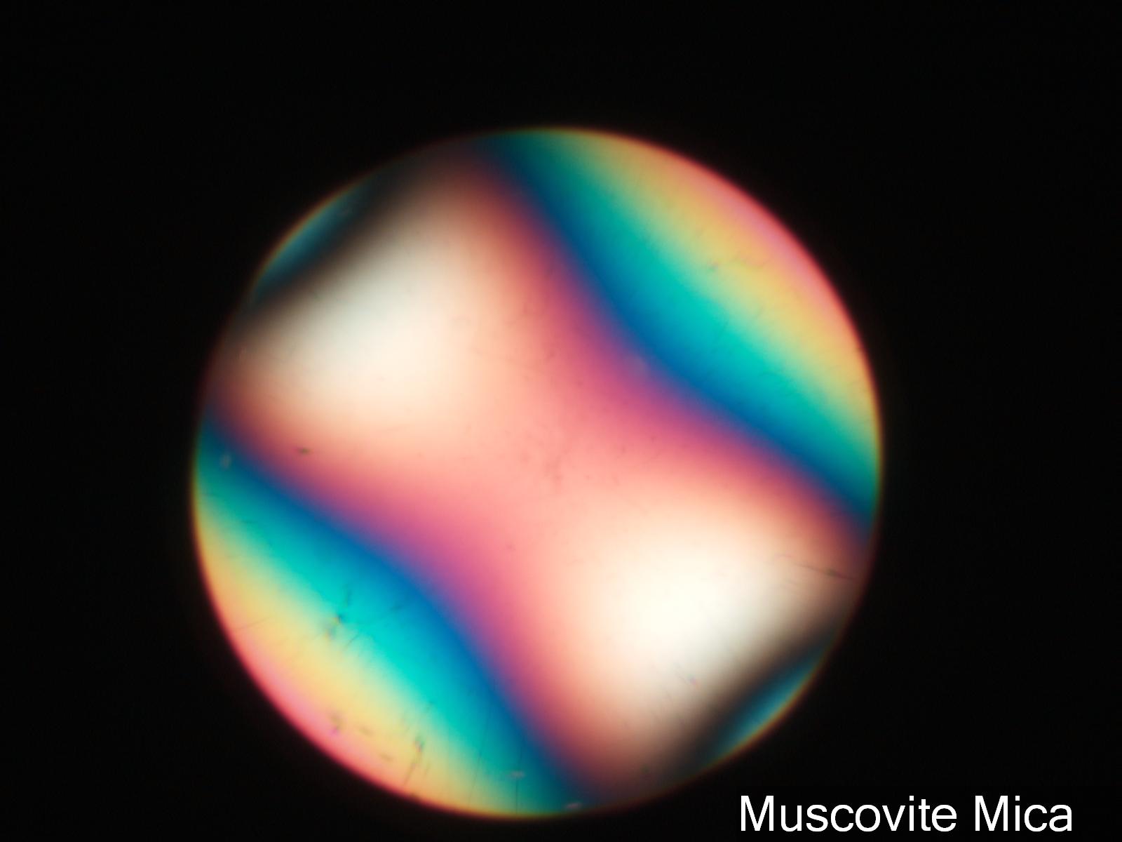
Biaxial Interference Figure, Muscovite Mica
The crystal in this view has been rotated 45 degrees from its darkest position (extinction Position). The optical axes here are in the second and fourth quadrant.
Transmitted Cross Polarized Light, Conoscopic View
Fleischer, Michael, Ray E. Wilcox, and John J. Matzko, THE MICROSCOPIC DETERMINATION OF THE NONOPAQUE MINERALS,U.S. Geological Survey Bulletin 1627, 1984.
Definition/Function:
The acute bisectorix for most of the mica minerals is normal to the main cleavage plane. As a result, the biaxial interference image can be seen when the mica flake is lying flat on the microscope slide. The biaxial interference figure changes dramatically as the stage is rotated, unlike the uniaxial interference figure. In its extiction position the center of the field is dominated by a relatively wide dark band. Rotated off of this position two hyperbola centered on the two optic axis become visible if the numerical apurature of the objective is large enough and the view is down the acute bisectorix.Significance in the Environment:
The interference figure can be used to determine the crystal group that the particle belongs to, uniaxial or biaxial, the optical sign of the mineral, and the orientation of the key refractive indices of the mineral. In the case of a biaxial crystal the Beta refractive index is oriented normal to the line conecting the two optic axes. The grain can be oriented and the polarizing filter that aligns with the Beta direction can be left in and the Beta refractive index can be measured using different refractive index oils. With the crystal class (biaxial), the opitcal sign (negative in the case of mica), and the Beta refractive index the mineral can be search in any tablular reference for mineral identification. Another key property of the biaxial minerals is the angle between the two optical axes, the 2V angle. As the angle increases the distance between the projection of the optical axes increases in the interference figure. Muscovite mica has a large 2V angle but biotite mica has a 2V angle of only a few degrees. The optical axes are near the egde of the field of view for an objective with a numerical apurature of 0.65 for muscovite but are near the center of the field of view for biotite.Characteristic Features:
The biaxial interference figure, using linear polarizing filters, consists of the two melatopes (projection of the optical axes), the isogyres (dark band associated with each melatope), and the isochromes (colored bands).References:
http://www.microscopy-uk.org.uk/mag/artsep03/dwmica.html (An enjoyable adventure discovering mica in the environment and exploring with the microscope)Fleischer, Michael, Ray E. Wilcox, and John J. Matzko, THE MICROSCOPIC DETERMINATION OF THE NONOPAQUE MINERALS,U.S. Geological Survey Bulletin 1627, 1984.
