
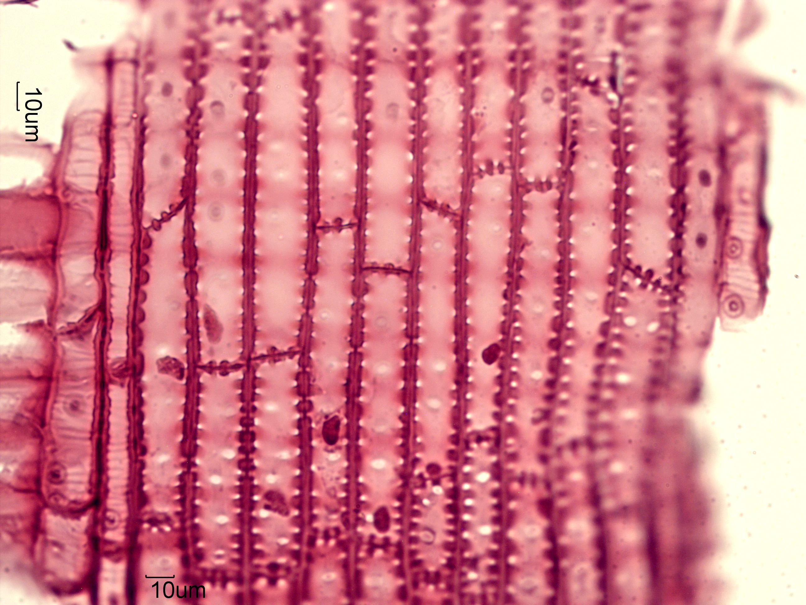
Douglas Fir, Radial-Section
This is an image from a stained standard section of Pseudotsuga menziesii made by John P. Limbach of Ripon, Wisconsin. This shows the nodular walls and termination of the vertical parenchyma. They don't contain the helical thickening seen in the vertial tracheids. The pits in the tracheids tend to be borderless.
Transmitted Brightfield Illumination
http://en.wikipedia.org/wiki/Douglas-fir
Definition/Function:
KINGDOM: Plantae DIVISION: Pinophyta CLASS: Pinopsida ORDER: Pinales FAMILY: Pinaceae GENUS: Pseudotsuga SPECIES: menziesiiSignificance in the Environment:
Characteristic Features:
Associated Particles:
References:
Core, H.R., W.A. Cote, and A.C. Day, WOOD STRUCTURE AND IDENTIFICATION, Syracuse Wood Science Series, Vol. 6, 1979.http://en.wikipedia.org/wiki/Douglas-fir
