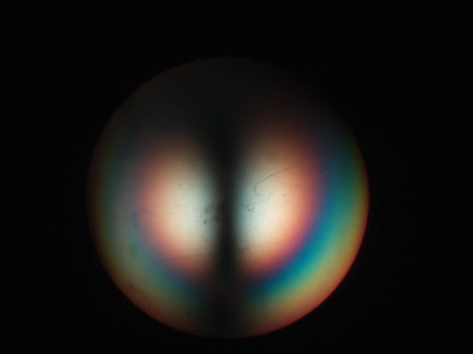
Biaxial Interference Figure - Sucrose (sugar)
The crystal in this view is in its darkest position
(extinction Position). The optical axes here are on the vertical
line runing through the center of the image. The dark band running horizontally
blots out the upper part of the image.
Transmitted Cross Polarized Light, Conoscopic View
Definition/Function:
Many of the sucrose crystals in granulated sugar are in a position that allows an
optical axis to nearly an acute bisectorix view. The biaxial
interference figure changes dramatically as the stage is rotated, unlike the uniaxial
interference figure. In its extiction position the center
of the field is dominated by a relatively wide dark band. Rotated off of this position
normally only one of the two isogyres centered on the
two optic axis are visible.
Significance in the Environment:
The interference figure can be used to determine the crystal group that the particle
belongs to, uniaxial or biaxial, the optical sign of the
mineral, and the orientation of the key refractive indices of the mineral. In the case
of a biaxial crystal the Beta refractive index is
oriented normal to the line conecting the two optic axes. The grain can be oriented and
the polarizing filter that aligns with the Beta direction
can be left in and the Beta refractive index can be measured using different refractive
index oils. With the crystal class (biaxial), the opitcal
sign (negative in the case of mica), and the Beta refractive index the mineral can be
search in any tablular reference for mineral identification.
Another key property of the biaxial minerals is the angle between the two optical axes,
the 2V angle. As the angle increases the distance
between the projection of the optical axes increases in the interference figure.
Muscovite mica has a large 2V angle but biotite mica has a 2V
angle of only a few degrees. The optical axes are near the egde of the field of view for
an objective with a numerical apurature of 0.65 for
muscovite but are near the center of the field of view for biotite.
Characteristic Features:
The biaxial interference figure consists of the two melatopes (projection of the optical
axes), the isogyres, and the isochromes (colored bands).
References: