
Weld Debris
Transmitted Off Circular Polarized Light Illumination
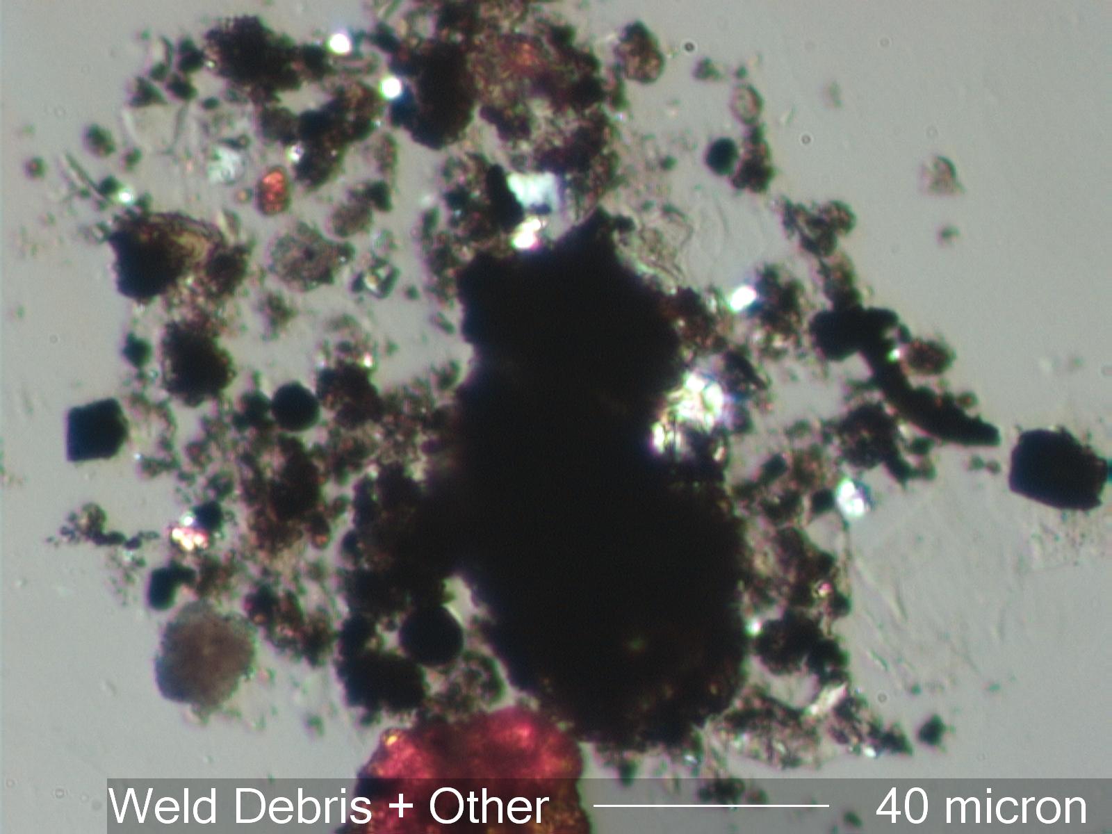
Welding and associated activities are represented in this photograph. Rust is present at various levels of hydration. It can be seen as the yellow orange particle in the upper left and as the reddish orange large particle at the lower center of of the photograph. The characteristic magnetite spheres can be seen as the black circular outlines with bright edges in this image. Their identification should be confirmed with reflected darkfield illumination. If they are magnetite spheres then there will be a bright circular reflection near the center of the particle, which is the bright ring of light coming from the reflected light darkfield illuminator. The fact that the reflected light in the magnetite sphere is circular confirms the spherical shape of the particle. Emery abrasive particles are present here as well as the abraded metal particles created by abrasive grinding or shaping. There are two emery particles particles visible as bright white objects extending from the dark center mass in this image. The abraded metal particles appear black with transmitted light and can only be identified with darkfield reflected light. The gray particle in the lower left is a flattened paint sphere. This was probably flattened in the process of pressing the tape used to collect this sample onto the surface. The fact that it flattens by splitting, which results in the four-lobed shape, indicates that the particle is held together by a tough outer film, the hardened vehicle of the paint. The other particles in the field include high iron weld slag (black irregular flakes), flux residues (brownish isotropic agglomerates), skin flakes (faint thin films at the lower right), and other common environmental particles.
Welding and associated activities are represented in this photograph. Rust is present at various levels of hydration. It can be seen as the yellow orange particle in the upper left and as the reddish orange large particle at the lower center of of the photograph. The characteristic magnetite spheres can be seen as the black circular outlines with bright edges in this image. Their identification should be confirmed with reflected darkfield illumination. If they are magnetite spheres then there will be a bright circular reflection near the center of the particle, which is the bright ring of light coming from the reflected light darkfield illuminator. The fact that the reflected light in the magnetite sphere is circular confirms the spherical shape of the particle. Emery abrasive particles are present here as well as the abraded metal particles created by abrasive grinding or shaping. There are two emery particles particles visible as bright white objects extending from the dark center mass in this image. The abraded metal particles appear black with transmitted light and can only be identified with darkfield reflected light. The gray particle in the lower left is a flattened paint sphere. This was probably flattened in the process of pressing the tape used to collect this sample onto the surface. The fact that it flattens by splitting, which results in the four-lobed shape, indicates that the particle is held together by a tough outer film, the hardened vehicle of the paint. The other particles in the field include high iron weld slag (black irregular flakes), flux residues (brownish isotropic agglomerates), skin flakes (faint thin films at the lower right), and other common environmental particles.
