|
Resolution vs Visibility Through the Microscope
The resolution of objects viewed through the light microscope is a calculated
value based on configuration of the microscope.
Visibility is a function of contrast. To take advantage of resolution there must
be sufficient contrast to see the subject
but objects far below the resolution limit of a specific microscope
configuration can be made visible with sufficient
contrast. This is the same phenomena that allows us to see the stars in the
night sky, all of which are far below the
resolution limit of our eyes. A few examples through the microscope are provided
below showing methods of improving the
resolution and/or visibility of objects viewed through the "standard" light
microscope. Click on the photographs below for
more information.
Resolution and Ocular Magnification
The resolution of the microscope is determined by the objective and the
condenser. Numerical apurture is important but the diffraction
maxima collected at the back focal plane of the objective is more important. The
eyepiece must provide sufficient magnification for the
observer to see what is being resolved. The images below provide a simple
example.
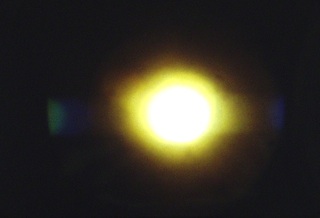
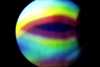
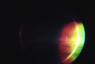
These two images show the diffraction maxima at the back focal plane of the
10X objective when viewing a diffraction grating with a
spacing of 1.9 micrometers. The first is using brightfield illumination. The
"0" order maximum at the center provides no information
on spacing of the grating. Only higher order maxima at the very edge of the
field of view, blue and green in this example, indicate
resolving power. The second is using darkfield illumination. Higher order
diffraction maxima fill the back focal plane.
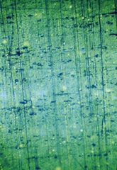
These two images show the effect of the eyepiece magnification. The eyepiece
magnification in the first image is sufficient to see the
resolved grating. The eyepiece magnification in the second image is not
sufficient for the camera to detect the resolved grating.
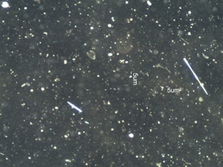
Brightfield vs Darkfield Illumination

Brightfield vs Oblique Illumination
TN.jpg)
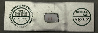
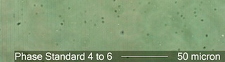
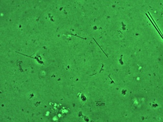
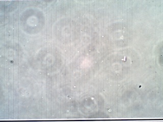
Phase Contrast vs Visibility Through the Microscope
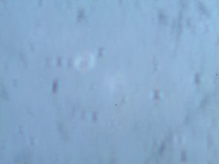
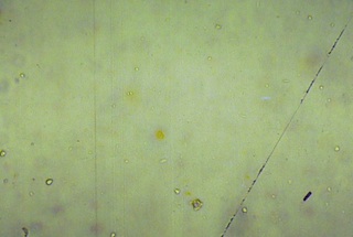
Phase contrast microscopy makes "phase" objects more visible. Every object is a
"phase" object strictly speaking but some objects are
primarily phase objects, being nearly invisible using standard brightfield
illumination. Phase constrast does not always improve
visibility. Small objects with significantly different refractive indices than
the medium in which they are mounted will be visible using
darkfield illumination even when they are invisible using phase contrast. The
"Phase Contrast Test Slide" is a good example, as shown
below.
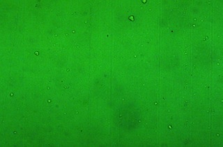
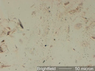
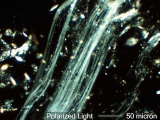
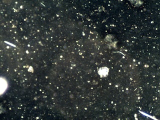
Polarized Light vs Visibility Through the Microscope
Objects with more than one refractive index can be made self-luminous by using
crossed polarized light. This can create sufficient contrast to see the
object.
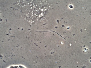
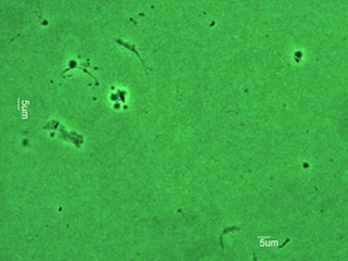
Asbestos Fibers and Visibility Through the Light Microscope
Much has been made of the need for transmission electron microscopy (TEM)
"because the light microscope can't see
asbestos fibers thinner than 0.25 micrometers". This quote is in error on many
counts, but is a common
misconception. First, it is not the microscope that "sees", but rather the human
eye establishes the detection limit.
If the light source is not "on" the resolution limit for the microscope has not
changed but the eye won't see anything.
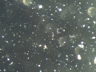
Second, the ability to "see" (detect) an object is not dependent on resolution.
This point has been made for the general
case above but will be made again here in the specific case of asbestos fibers.
Third, after over thirty years of
diligent searching there has been no evidence that asbestos fibers shorter than
five micrometers cause any health effect
more detrimental than nuisance dust. Fresh crystalline silica is more
detrimental in this respect. That challenges the
"need" for an instrument that can "see" particles that are not apparently
detrimental as a replacement for light
microscopy. We will continue monitoring with the TEM because it is required in
many governement and industrial
regulations, but it is not a "superior" method in the case of hazardous asbestos
detection or identification at sites
of asbestos removal projects.
|